Skip to main content
W.M. Keck Center for Cellular Imaging
W.M. Keck Center for Cellular Imaging
Main menu
Jobs
Contact
Faculty & Staff
Former Staff
Students
Former Students
Workshop
General Information
Workshop 2026
Workshop 2025
Workshop 2025 Materials
Workshop 2024
Workshop 2023
Previous Workshops
Microscopy Facility
Equipment
Instructions
Techniques
Accessories
Internal Advisory Committee
Former Internal Advisory Committee Members
Software
Server
User Fees
User Support
Education
Microscopy Course
FRET Literature
FRET Microscopy
PERIASAMY LAB
Publications
Workshop 2026
FRET
- Any -
True
False
FLIM
- Any -
True
False
Clinical Application
- Any -
True
False
Microscopy
- Any -
True
False
Characterization of phototoxic effects in multiphoton FLIM
Proc. SPIE 11965, 119650B (3 March 2022) (2022)
Characterization of mitochondrial dysfunction due to laser damage by 2-photon FLIM microscopy
Sci Rep. 2022 Jul 13;12(1):1. (2022)
Investigation of metabolism in cancer specimens using Fluorescence Lifetime Imaging Microscopy
Asian Journal of Physics, March 2021 (2021)
Machine learning architecture to predict drug response based on cancer cell FLIM images
Proc. SPIE 11648,16481G (March 2021) (2021)
Cyclic compression increases F508 Del CFTR expression in ciliated human airway epithelium
Am J Physiol Lung Cell Mol Physiol. 2019 Aug 1; 317(2): L247–L258 (2019)
Single‐cell redox states analyzed by fluorescence lifetime metrics and tryptophan FRET interaction with NAD(P)H
Cytometry Part A. 2019, 95: 110-121. (2019)
Intraneuronal Tau Misfolding Induced by Extracellular Amyloid-β Oligomers
Journal of Alzheimer's Disease, vol. Pre-press, no. Pre-press, pp. 1-14 (2019)
A novel lysosome-to-mitochondria signaling pathway disrupted by amyloid-ß oligomers
The EMBO Journal 37: e100241 (2018)
Segmented cell analyses to measure redox states of autofluorescent NAD(P)H, FAD & Trp in cancer cells by FLIM
Sci. Rep. 8: 79 (2018)
Mechanical and signaling roles for keratin intermediate filaments in the assembly and morphogenesis of Xenopus mesendoderm tissue at gastrulation
The Company of Biologists Ltd (2017)
O-Aminobenzoyl-S-Nitrosoglutathione: a Fluorogenic, Cell Permeable, Pseudo-Substrate for S-Nitrosoglutathione Reductase
Free Radical Biology and Medicine 108: 445–451 (2017)
FLIM-FRET Image Analysis of Tryptophan in Prostate Cancer Cells
Proc SPIE (ECBO) 10414: 1041402-Pp1-5 (2017)
Effects of Anti-Cancer Drug Doxorubicin on Endogenous Biomarkers NAD(P)H, FAD & Trp in prostate cancer cells- a FLIM Study
Proc. of SPIE, Vol. 10069: 100691L-Pp1-6 (2017)
Mechanical and signaling roles for keratin intermediate filaments in the assembly and morphogenesis of Xenopus mesendoderm tissue at gastrulation
Development. 144(23):4363-4376 (2017)
Investigation of Mitochondrial Metabolic Response to Doxorubicin in Prostate Cancer Cells: An NADH, FAD and Tryptophan FLIM Assay
Sci. Rep. 7: 10451 (2017)
Augmentation of CFTR maturation by S-nitrosoglutathione reductase
AJP Lung Cellular and Molecular Physiology (2016)
Investigation of prostate cancer cells using NADH and Tryptophan as biomarker: multiphoton FLIM-FRET microscopy
Proc. SPIE Int. Soc. Opt. Eng.7712: 97120Q. pp1-5. (2016)
FLIM data analysis of NADH and tryptophan autofluorescence in prostate cancer cells
Proc. SPIE Int. Soc. Opt. Eng.9712: 97122E. pp 1-6. (2016)
Three‐color confocal Förster (or fluorescence) resonance energy transfer microscopy: Quantitative analysis of protein interactions in the nucleation of actin filaments in live cells
Cytometry A., PMID 25755111 (2015)
Confocal immunofluorescence FRET microscopy to investigate eNOS and GSNOR localization and interaction in pulmonary endothelial cells
Proc. SPIE Int. Soc. Opt. Eng. 9329: 93290G. (2015)
Localizing Protein–Protein Interactions in Living Cells Using Fluorescence Lifetime Imaging Microscopy
Methods in Mol. Biol., Vol. 1251: 83-108 (2015)
Microscopy Core Facilities: Results of an International Survey
Microscopy Today March: pp36-44 (2014)
Development of an AP-FRET Based Analysis for Characterizing RNA-Protein Interactions in Myotonic Dystrophy (DM1)
PLoS ONE 9(4):e95957 (2014)
Advanced Light Microscopy
Methods 15: 66(2):121-123 (2014)
Förster resonance energy transfer microscopy and spectroscopy to localize protein-protein interactions in live cells
Cytometry A. 83A(9): 780-793 (2013)
Investigation of tryptophan–NADH interactions in live human cells using three-photon fluorescence lifetime imaging and Förster resonance energy transfer microscopy
J. Biomed. Opt. 18(6): 060501 (2013)
IQGAP1 interactome analysis by In Vitro reconstitution and live cell 3-color FRET microscopy
Cytoskeleton, 70: 819-836 (2013)
Non-invasive in vivo imaging of breast cancer cell internalization of transferrin by near infrared FRET
PLoS ONE 8(11): e80269 (2013)
Monitoring Protein Interactions in Living Cells with Fluorescence Lifetime Imaging Microscopy
Meth. Enzymol., 504: 371-391 (2012)
Mouse primitive streak forms in situ by initiation of epithelial to mesenchymal transition without migration of a cell population
Wiley Online Library (2012)
Investigating protein-protein interactions in living cells using fluorescence lifetime imaging microscopy
Nature Protocol Vol. 6 No. 9 (2011)
FRET Microscopy in 2010: The Legacy of Theodor Förster on the 100th Anniversary of his Birth
ChemPhysChem,12:462-474 (2011)
Three-Color Spectral FRET Microscopy Localizes Three Interacting Proteins in Living Cells
Biophysical J. Vol. 99, 1274-1283 (2010)
Additional correction for energy transfer efficiency calculation in filter-based Förster resonance energy transfer microscopy for more accurate results
J Biomed. Opt. 15(2) (pp1-3) (2010)
FLIM Microscopy in Biology and Medicine
CRC Press (2009)
Characterization of an orange acceptor fluorescent protein for sensitized spectral fluorescence resonance energy transfer microscopy using a white-light laser
J. Biomed. Opt. 14(5) (2009)
PTK7 is essential for polarized cell motility and convergent extension during mouse gastrulation
Development 136: 2039-2048 (2009)
Quantitation of Protein–Protein Interactions: Confocal FRET Microscopy
Meth. Cell Biol. 89: 569-598 (2009)
Characterization of an improved donor fluorescent protein for Förster resonance energy transfer microscopy
J. Biomed. Opt. 13 (pp1-9) (2008)
Characterization of spectral FRET imaging microscopy for monitoring nuclear protein interactions
J Microscopy, 228:139-152 (2007)
Receptor Complexes Cotransported via Polarized Endocytic Pathways Form Clusters with Distinct Organizations
Mol. Biol. Cell. 18:2226-2243 (2007)
Localization of protein-protein interactions in live cells using confocal and spectral imaging FRET microscopy
Indian J Exp. Biol., 45(01):48-57 (2007)
Angiotensin II Type 2 Receptor–Bradykinin B2 Receptor Functional Heterodimerization
Hypertension (JAHA, Journal of the American Heart Association) 48:1-7 (2006)
Monitoring dynamic protein interactions with photoquenching FRET
Nature Methods 3(7):519-524 (2006)
Intensity Range Based Quantitative FRET Data Analysis to Localize Protein Molecules in Live Cell Nuclei
J. Fluorescence. 16:95-104 (2006)
Issues in confocal microscopy for quantitative FRET analysis
Microscopy research and Techniques. 69:196-206 (2006)
Molecular Imaging: FRET Microscopy and Spectroscopy
Academic Press (2005)
Imaging protein molecules using FRET and FLIM microscopy
Current Opinion in Biotechnology. 16:19-27 (2005)
Characterization of two‐photon excitation fluorescence lifetime imaging microscopy for protein localization
Microscopy Research and Techniques. 63:72-80 (2004)
Illuminating protein interactions in tissue using confocal and two-photon excitation fluorescent resonance energy transfer microscopy
J. Biomed. Opt. 8: 347-356 (2003)
One- and two-photon fluorescence resonance energy transfer microscopy to establish a clustered distribution of receptor-ligand complexes in endocytic membranes
J Biomed. Opt. 8(3), 339-346 (2003)
Fluorescence resonance energy transfer (FRET) microscopy imaging of live cell protein localizations
The Journal Of Cell Biology, Volume 160, Number 5, March 3, 2003 629-633 (2003)
Protein localization in cells and tissues using FLIM and FRET
Differentiation. 71:528-541 (2003)
Imaging The Localized Protein Interactions Between Pit-1 And The CCAAT/Enhancer Binding Protein Alpha (C/EBP?) In The Living Pituitary Cell Nucleus
Day Et Al. Revised ME 02-0136 (2003)
Imaging the localized protein interactions between Pit-1 and the CCAAT/enhancer binding protein alpha (C/EBPα) in the living pituitary cell nucleus
Mol. Endo. 17(3) (2003)
Confocal FRET Microscopy To Measure Clustering Of Ligand-Receptor Complexes In Endocytic Membranes
Biophysical Journal 85: 559-571 (2003)
Dynamic Imaging Using Fluorescence Resonance Energy Transfer
Biotechniques Vol. 32 No. 6 (2002)
Nanosecond fluorescence resonance energy transfer‐fluorescence lifetime imaging microscopy to localize the protein interactions in a single living cell
J Microscopy. 205:3-14 (2002)
Fluorescence Lifetime Imaging (FLIM) Of Green Fluorescent Fusion Proteins In Living Cells
Methods In Molecular Biology, Vol. 183: Green Fluorescent Protein: Applications And Protocals (2002)
Characterization Of One- And Two-Photon Excitation Fluorescence Resonance Energy Transfer Microscopy
Methods In Press (December, 2002) (2002)
Survival of bundleless hair cells and subsequent bundle replacement in the bullfrog's saccule
J. Neurobiol.50, 81-92 (2002)
Truncated Estrogen Receptor Product-1 Suppresses Estrogen Receptor Transactivation by Dimerization with Estrogen Receptors α and β
J. Biol. Chem., 275: 7158-7166 (2002)
Methods in Cellular Imaging
Oxford University Press (2001)
Fluorescence Resonance Energy Transfer Microscopy Of Localized Protein Interactions In The Living Cell Nucleus
Methods 25, 4-18 (2001)
Fluorescence Resonance Energy Transfer Microscopy: A Mini Review
Journal Of Biomedical Optics 6(3) 28-291 (July 2001) (2001)
Fluorescence Microscopy Study of Heterogeneity in Polymer-supported Luminescence-based Oxygen Sensors
Microscopy and Microanalysis. 6: 551-556 (2000)
Error Analysis Of The Rapid Lifetime Determination Method For Double-Exponential Decays And New Windowing Schemes
Anal. Chem.1999, 71,947-952 (1999)
An Evaluation of Two-Photon Excitation Versus Confocal and Digital Deconvolution Fluoescence Microscopy Imaging in Xenopus Morphogenesis
Microscopy Research and Technique 47:172-181 (1999)
Fret Imaging Of Pit-1 Protein Interactions In Living Cells
Journal Of Biomedical Optics 3(2), 154-160 (April 1998) (1998)
Visualizing Protein Interactions In Living Cells Using Digitized GFP Imaging And FRET Microscopy
Methods In Cell Biology, Vol. 58 (1998)
Time-Resolved Fluorescence Lifetime Imaging Microscopy Using A Picosecond Pulsed Tunable Dye Laser System
Rev. Sci. Instrum. 67 (10), October 1996 (1996)
High-Speed Fluorescence Microscopy: Lifetime Imaging in the Biomedical Sciences
J. Micros. Soc. Am., 1: 13-23 (1995)
Computerized fluorescence microscopic vision in the biomedical sciences
J. Comp. Assist. Micros., 6: 1-26 (1994)
Detection Of Human Papillomavirus Type 16/18 DNA In Cervicovaginal Cells By Fluorescence Based In Situ Hybridization And Automated Image Cytometry
Cytometry 15:245-257 (1994)
Fluorescence Lifetime Imaging Microscopy (FLIM): Instrumentation and Applications
Critical Rev. Analyt. Chem., 23(5): 369-395 (1992)
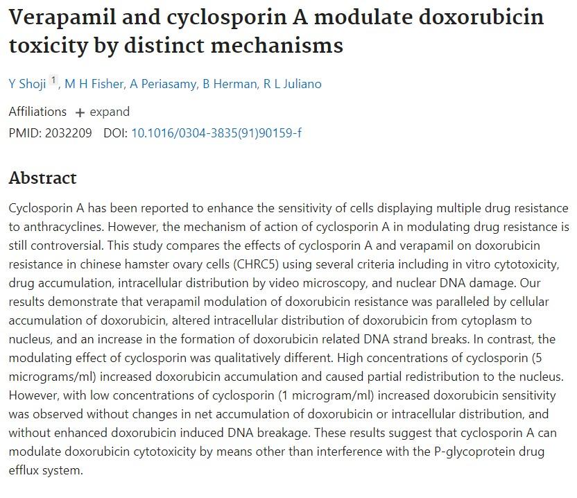
Verapamil and cyclosporin A modulate doxorubicin toxicity by distinct mechanisms
Cancer Letters. 57: 209-218 (1991)