Leica STELLARIS 8 tauSTED Super Resolution microscopy system [Funded by NIH OD 030409; 2022]
This system equipped with an inverted microscope, coupled to a confocal, FLIM (FALCON), and tauSTED microscopy system.
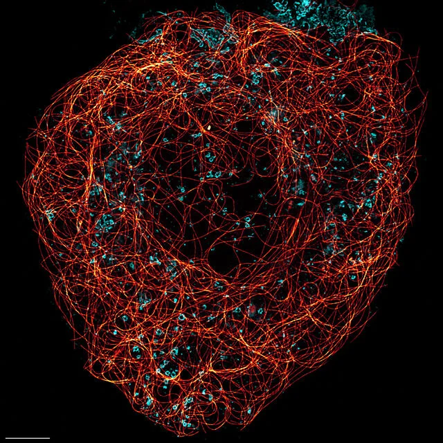
The Leica STELLARIS 8 tauSTED (Stimulated Emission Depletion) is a fully integrated system with FALCON (FAst Lifetime CONtrast) imaging, providing fast, intuitive access to structural details inside the sample, far beyond the diffraction limit. The tauSTED microscope is based on a Leica STELALRIS 8 spectral confocal scan head. The system has unique features that allow for unsurpassed flexibility and sensitivity in confocal, lifetime (FLIM), and STED imaging microscopy.
The lateral resolution of Leica tauSTED is about 20-30 nm for fixed specimen and <70 nm for live specimens. The Z resolution is about 200nm.
tauSTED Depletion lines - The tauSTED configuration has three STED depletion laser lines: 592 nm, 660 nm, and 775 nm to cover the complete visible spectrum for the far-red fluorophores. The notch filter set allows to block the excitation and depletion light in order to improve the efficiency of the fluorescence emission signal. As mentioned in the confocal below, the WLL laser lines (440-790 nm) are required to excite labeled fluorophores to acquire tauSTED images.
Leica STELLARIS 8 Laser scanning Confocal/spectral imaging microscopy system [Funded by NIH-OD 030409; 2022]
Leica STELLARIS 8 confocal system equipped with pulsed white light laser (WLL; repetition rate 78MHz) and tunable excitation wavelengths from 440 nm to 790 nm [Laser power per line: > 0.9 mW 440-485 nm, > 1.8 mW 485-790 nm; if more power is required more lines (up to 8 lines) can be used simultaneously]. This WLL will be used in both, confocal and tauSTED mode. In addition, the system will include a 405 nm excitation laser line in a much-needed spectral range not covered by the WLL.
The enhanced detection efficiency and the optimal match of excitation and detection enable long time live cell imaging. The combination of Field-of-View (FOV) or Tandem scanner with rolling average and LIGHTNING delivers superb image quality at full speed and high resolution (120-140 nm) with better SNR (signal to noise ratio). The tandem scanner (resonant scanner @16KHz) provides 28 frames/second (fps) @512 x 512 and up to 290 fps @512 x 16.
Leica FALCON One-photon TD FLIM microscopy system [Funded by NIH-OD 030409; 2022]
The FALCON, based on time correlated single photon counting (TCSPC) provides fast acquisition, processing, and analysis of FLIM and real time display of the FLIM and tauSTED images. The acquisition software, fully integrated with LAS X, enables FLIM for any imaging workflow (time lapse, 3D, lambda scan, combination with mosaic imaging). It drives the IRF (instrument response function) and includes n-exponential Reconvolution and n-Exponential Tail fitting with up to 5 components, dead time correction and incomplete decay fitting algorithm. The FLIM analysis software is essential to process the tauSTED images. FLIM analysis software can also be used on ROIs as well as on a series of images. This FALCON FLIM is ideal for time-lapse FLIM
Components & Features
- Automated stage (for screening and tile imaging) and focus.
- Temperature controlled Chamber
- Objective lenses: 5x/0.15, 10x/0.40, 20x/0.75, 20x/0.8 multi-immersion, 40x/1.3 oil, 63x/1.4 oil, 100x/1.4 oil.
- For tauSTED imaging 100x/1.4 oil and 86x/1.2W and this is a Plan apochromatic water immersion objective lens with optimal chromatic correction and transmission for 2D/3D nanoscopy of live and fixed samples in aqueous media.
- Excitation lasers: 405 nm; pulsed white light laser (WLL; repetition rate 78MHz) and tunable excitation wavelengths from 440 nm to 790 nm.
- For tauSTED depletion laser lines: three STED depletion laser lines: 592 nm, 660 nm, and 775 nm to cover the complete visible spectrum for the far-red fluorophores.
- Internal Detectors: The AOBS in combination with prisms generates superb fluorophore emission signals to the detectors. The system also includes 5 independent detection channels, two HyD detector channels with a photon detection efficiency (PDE) of ~58% @500nm and another two HyDs, where efficiency is ~46% @500nm; the 5th channel HyD has ~26% efficiency at 635 nm for far red detection.